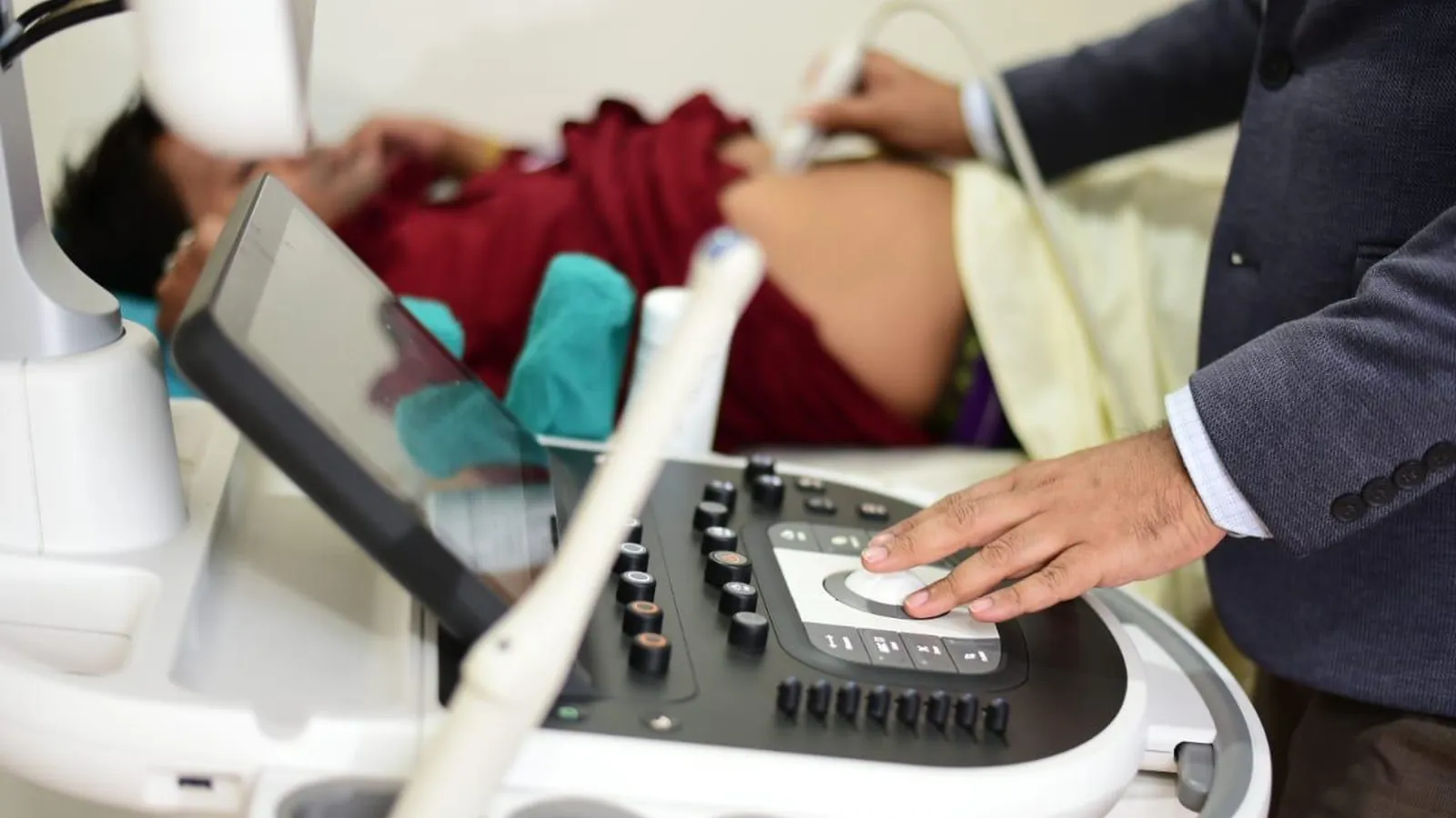
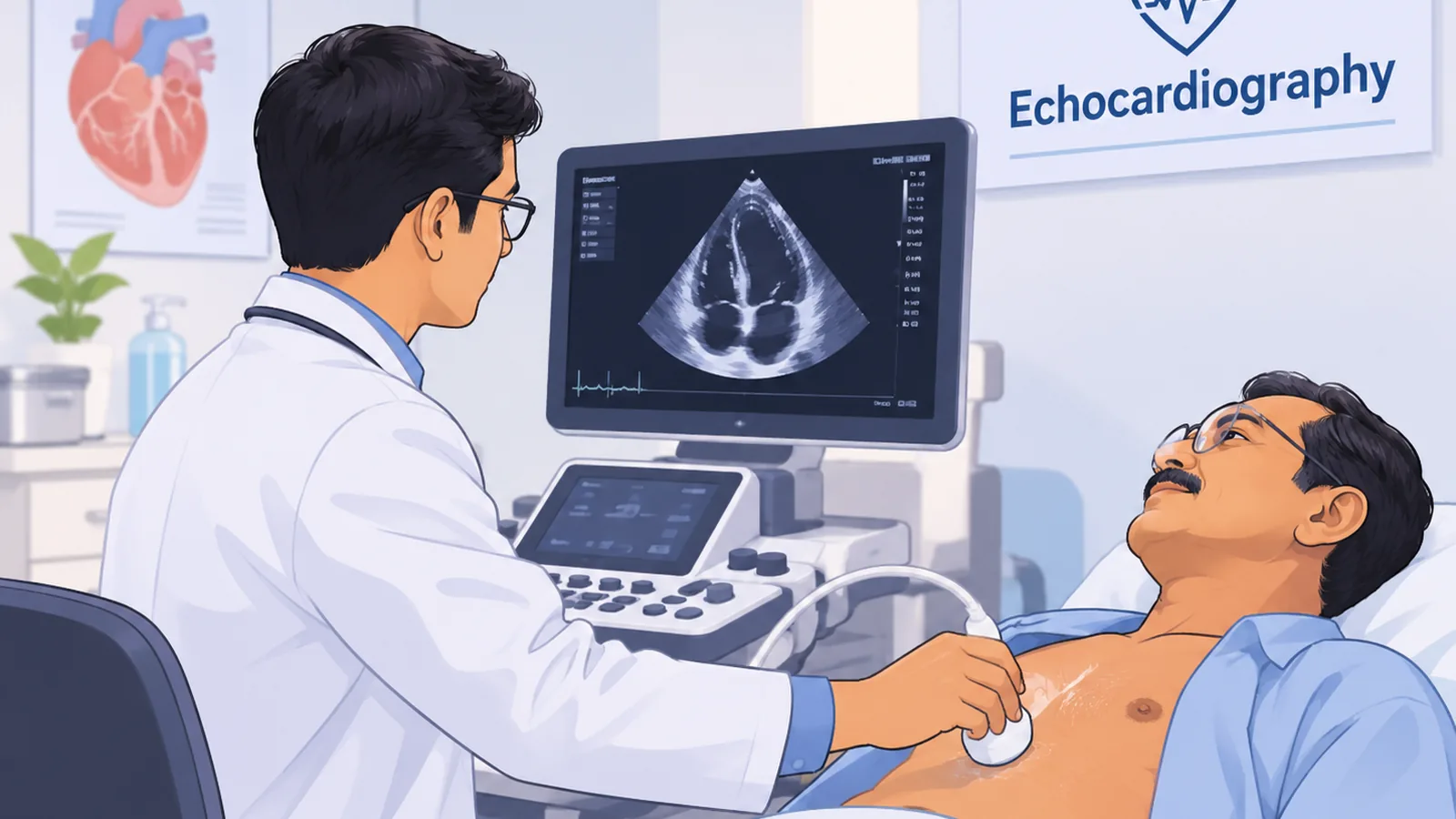
An echocardiogram, or echo, is an ultrasound of the heart. A probe is moved across the chest and the screen shows the heart beating in real time: the chambers filling and emptying, the valves opening and closing. There is no radiation, no injection, and no pain. Most people are more nervous about the report than the scan, because echo reports are full of terms that sound worse than they are. This article explains what the test shows, what it does not, and how to make sense of the result. UHS Vista offers echocardiography in Guwahati at its South Sarania centre, as part of its imaging services.
What an Echo Measures
An ECG records the electrical signal that makes the heart beat. An echo shows the physical heart. It measures the size of each chamber, the thickness of the walls, how well the muscle squeezes, how the four valves behave, and whether there is fluid around the heart. It can also estimate the pressure in the blood vessels of the lungs, which rises in several heart and lung conditions.
The Terms You Will See on Your Report
Ejection fraction (EF or LVEF). The percentage of blood the left ventricle pumps out with each beat. A normal EF is roughly 55 to 70 percent. A reduced EF suggests the heart muscle is weak, which is the basis for diagnosing heart failure and judging how well treatment is working. A normal EF does not mean the heart is perfectly healthy, because some forms of heart failure occur with a normal EF.
Regurgitation (MR, TR, AR). A valve that does not close fully and lets a little blood leak backwards. Trivial or mild regurgitation, especially of the tricuspid or mitral valve, is seen in many healthy people and usually needs nothing more than awareness. Moderate or severe regurgitation is graded differently and is followed up.
Stenosis. A valve that does not open fully. This is more significant and is commonly linked in India to rheumatic heart disease, which most often affects the mitral valve.
Left ventricular hypertrophy (LVH). Thickening of the heart wall, often a result of long-standing high blood pressure.
Diastolic dysfunction. The heart is stiffer than normal when it relaxes and fills. Mild grades are common with age and high blood pressure.
Regional wall motion abnormality (RWMA). A part of the heart wall that moves less than the rest, which can suggest an old heart attack or reduced blood supply to that area.
Pericardial effusion. Fluid between the heart and its outer lining. Small amounts can be harmless, and larger amounts need investigation.
When Doctors Recommend an Echo
A cardiologist usually orders an echo to answer a specific question. Common reasons are breathlessness that is new or out of proportion to activity, swelling in the legs, a murmur heard through the stethoscope, fainting, palpitations that need an explanation, and long-standing high blood pressure or diabetes with suspected effects on the heart. It is also used to follow known conditions, to assess the heart before certain surgeries, and in children when a murmur or symptoms raise the question of a heart defect present since birth. Many murmurs in children turn out to be harmless, and the echo is what confirms that.
What an Echo Cannot Do
This is the point people most often misunderstand. A standard resting echo looks at the heart's structure and pumping, but it does not directly show blockages in the coronary arteries. Someone with significant blockages can have a normal echo at rest. If your doctor suspects coronary artery disease, tests such as a treadmill test, a stress echo, a CT scan of the coronary arteries, or an angiogram may be needed. A normal echo is reassuring about valves and muscle function. It is not a clearance certificate for the arteries.
Echo, ECG, and Treadmill Test
The ECG is quick and cheap, and it is the first test for rhythm problems and for a heart attack in progress. The echo answers structural questions. The treadmill test looks for signs of reduced blood flow to the heart during exertion. They answer different questions, and your cardiologist chooses based on your symptoms. If chest discomfort is what brought you here, our guide on chest pain that is not a heart attack explains how doctors tell the difference.
What Happens During the Scan
You lie on your left side or back with your chest uncovered, and gel is applied so the probe makes good contact. The technician takes images from several positions, and you may be asked to hold your breath briefly or turn onto your side. The scan typically takes 20 to 40 minutes. No fasting is usually needed, but check when booking. A loose, front-opening top makes things easier.
Frequently Asked Questions
Is an echo painful or harmful?
No. It uses sound waves, with no radiation or injections, and can be repeated safely.
Do I need to fast or stop my medicines?
Usually not. Confirm when you book, and carry a list of your current medicines for the doctor.
Can an echo detect a heart attack or blockage?
It can show damage from an earlier heart attack, such as a wall that moves poorly, but it does not directly show blockages in the arteries. A heart attack in progress is an emergency that needs immediate hospital care.
Can children have an echo?
Yes, and it is the standard way to check a heart murmur in a child. The scan is painless, though young children may need to be kept calm and still.
When Not to Wait
Sudden severe chest pain, pain spreading to the arm, jaw, or back, or chest discomfort with sweating, nausea, or breathlessness may be a heart attack. Go to the nearest emergency department immediately instead of booking a scan.
Echocardiography at UHS Vista
Echo and Doppler services are available at our South Sarania centre, alongside cardiology consultations. Bring any earlier ECG, echo, or blood reports so the doctor can compare them with the new scan. You can meet our cardiologists or book a scan.
Visit us: Dwarka Centre, Saniram Bora Road, behind Bora Service, South Sarania, Lachit Nagar, Guwahati, Assam
Timings: Monday to Saturday, 7:30 AM to 8 PM. Sunday, 7:30 AM to 1:30 PM.
Contact: +91 92879 88515 | uhsvista@gmail.com