
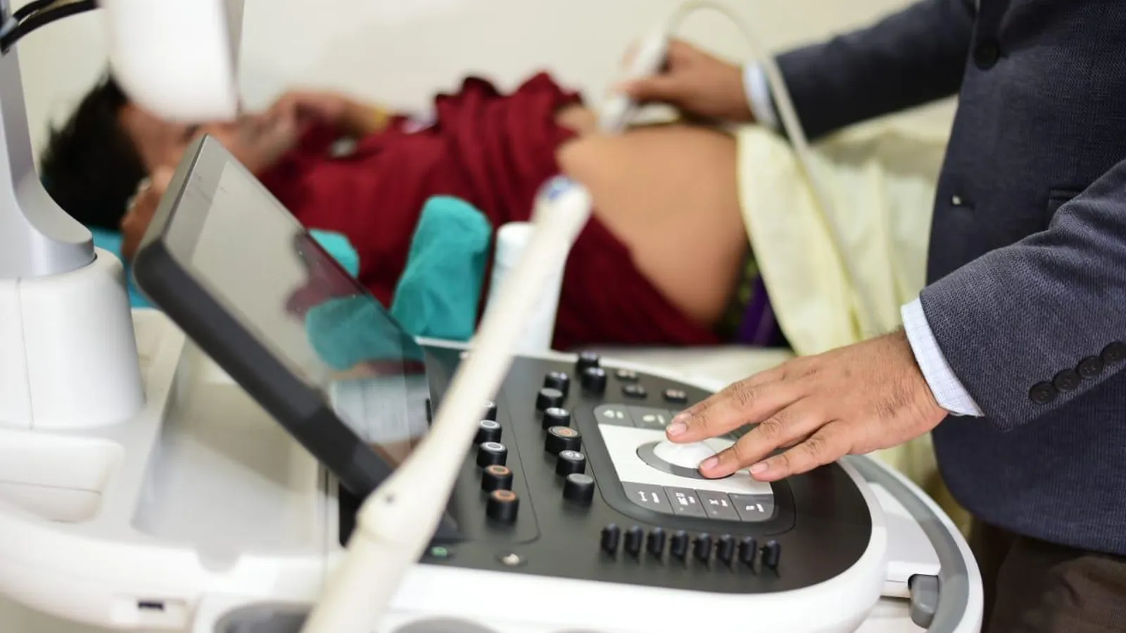
Most pregnancies involve several ultrasound scans, and each is timed for a reason. A scan done at the wrong week can miss what it was meant to find, or find something that needs no action. Knowing what each scan is for, and what it cannot do, makes the appointments less stressful. Your gynaecologist sets your schedule, so treat the timings below as the usual pattern and not a fixed rule. UHS Vista offers pregnancy ultrasound in Guwahati at its South Sarania centre, along with gynaecology consultations.
The Early Scan: Around 6 to 9 Weeks
The first scan answers basic but important questions: whether the pregnancy is inside the uterus, whether there is a heartbeat, and how many babies there are. A heartbeat is usually visible from about 6 weeks. At this stage the scan is often done internally (transvaginally), because the baby is too small to see well through the abdomen, and it is the way to rule out an ectopic pregnancy when there is pain or bleeding.
The early scan also fixes the due date. In the first trimester babies grow at almost the same rate, so measuring the crown-to-rump length dates the pregnancy to within about 5 to 7 days. That is more accurate than counting from the last period, which many women cannot place exactly. The due date set here is normally kept for the rest of the pregnancy, and every later growth measurement is judged against it.
The NT Scan: 11 to 14 Weeks
The nuchal translucency (NT) scan measures a small pocket of fluid at the back of the baby's neck. A thicker measurement, commonly 3.0 mm or more, is associated with a higher chance of conditions such as Down syndrome, and with some heart and structural problems. It is usually combined with a blood test (free beta-hCG and PAPP-A) and the mother's age to give a risk figure, such as 1 in 1,000 or 1 in 80.
This is screening, not diagnosis. A high-risk result does not mean the baby has a condition. It means further testing is worth discussing: a non-invasive blood test that analyses fetal DNA in the mother's blood, or a diagnostic procedure such as CVS or amniocentesis, which carry a small risk of miscarriage and are offered only after counselling. A low-risk result lowers the chance but cannot rule it out entirely.
The Anomaly Scan: 18 to 22 Weeks
This is the detailed scan, and it takes longer than the others. The sonographer examines the baby's brain, face, spine, heart, stomach, kidneys, abdominal wall, and limbs, and also checks the placenta, the amniotic fluid, and the number of vessels in the umbilical cord. The cervical length is often measured as well, since a short cervix can signal a higher risk of early labour.
Two findings cause the most worry and need explaining. A low-lying placenta seen at 20 weeks usually moves upward as the uterus grows, and it is rechecked later, around 32 to 34 weeks. And some soft markers, like a tiny bright spot in the heart, are often normal variants. Your doctor will tell you when a finding matters and when it does not.
The anomaly scan finds many structural problems but not all. Some conditions, including certain heart defects, are hard to see, and others only appear later in pregnancy. A normal scan is reassuring, though it is not a guarantee.
Growth Scans: Third Trimester
Growth scans estimate the baby's weight from measurements of the head, abdomen, and thigh bone, and compare them with expected values for the gestational age. A baby below the 10th percentile is described as small for gestational age. Many of these babies are simply small and healthy, and others are not growing as they should because of placental problems, so the doctor looks at the trend over several scans and at blood flow before drawing conclusions. The scan also checks the amount of amniotic fluid, the baby's position, and where the placenta lies.
Where Doppler Fits In
Doppler ultrasound assesses blood flow. A study of the uterine arteries around 20 to 24 weeks helps identify women at higher risk of pre-eclampsia. Later in pregnancy, Doppler of the umbilical artery and other vessels is used when a baby is small or when the mother has high blood pressure or another risk factor, to see whether the placenta is delivering enough. It is not needed in every pregnancy.
Is Ultrasound Safe?
Ultrasound uses sound waves, not radiation, and has no known harmful effects on the baby when done for medical reasons by trained staff. More scans are not automatically better. The number should follow your doctor's judgement about your pregnancy.
About Knowing the Baby's Sex
Under India's PCPNDT Act, determining or disclosing the sex of a baby is illegal. Scans are done to check the health of the baby and the pregnancy.
Preparing for a Scan
Early scans may require a comfortably full bladder, and the centre will tell you when you book. Later scans need no preparation. Wear a loose two-piece outfit, and bring earlier reports and your antenatal records so the doctor can compare measurements.
When to Call Your Doctor Instead of Waiting
Vaginal bleeding, severe or constant abdominal pain, leaking of fluid, a severe headache with blurred vision or sudden swelling of the face and hands, or noticeably reduced baby movements in later pregnancy should be assessed promptly. Do not wait for the next scheduled scan.
Frequently Asked Questions
How many ultrasounds will I have in pregnancy?
It varies. A typical low-risk pregnancy has an early scan, an NT scan, an anomaly scan, and often one or more growth scans. Your doctor may add scans if there is a reason.
Do I need a full bladder?
For early scans, usually yes. Later scans generally do not need it. The centre will tell you when you book.
Can the scan tell me if it is a boy or a girl?
No. Disclosing the sex of the baby is prohibited under the PCPNDT Act.
What if a scan finds something unusual?
Many findings are normal variants or need only a repeat scan. Your gynaecologist will explain what it means and whether further tests are needed.
Pregnancy Scans at UHS Vista
Ultrasound, Doppler, and gynaecology consultations are available at our South Sarania centre, so you can have your scan and discuss the result with the doctor on the same visit. See all imaging services or book an appointment.
Visit us: Dwarka Centre, Saniram Bora Road, behind Bora Service, South Sarania, Lachit Nagar, Guwahati, Assam
Timings: Monday to Saturday, 7:30 AM to 8 PM. Sunday, 7:30 AM to 1:30 PM.
Contact: +91 92879 88515 | uhsvista@gmail.com